We provide one-on-one training, method development, proposal writing, troubleshooting,
and data analysis services to help you meet your biofilm-related bioimaging needs.
Our staff are highly trained and specialized experts with Ph.D.'s in biofilm science.
Our Bioimaging and Analytical Core labs are equipped with state-of-the-art technologies
and methods to provide you with the best results and images of your biofilm or other
complex samples.
Acknowledgment of core facility services is required in publications. Please visit this page to learn how to properly acknowledge the Bioimaging Core.
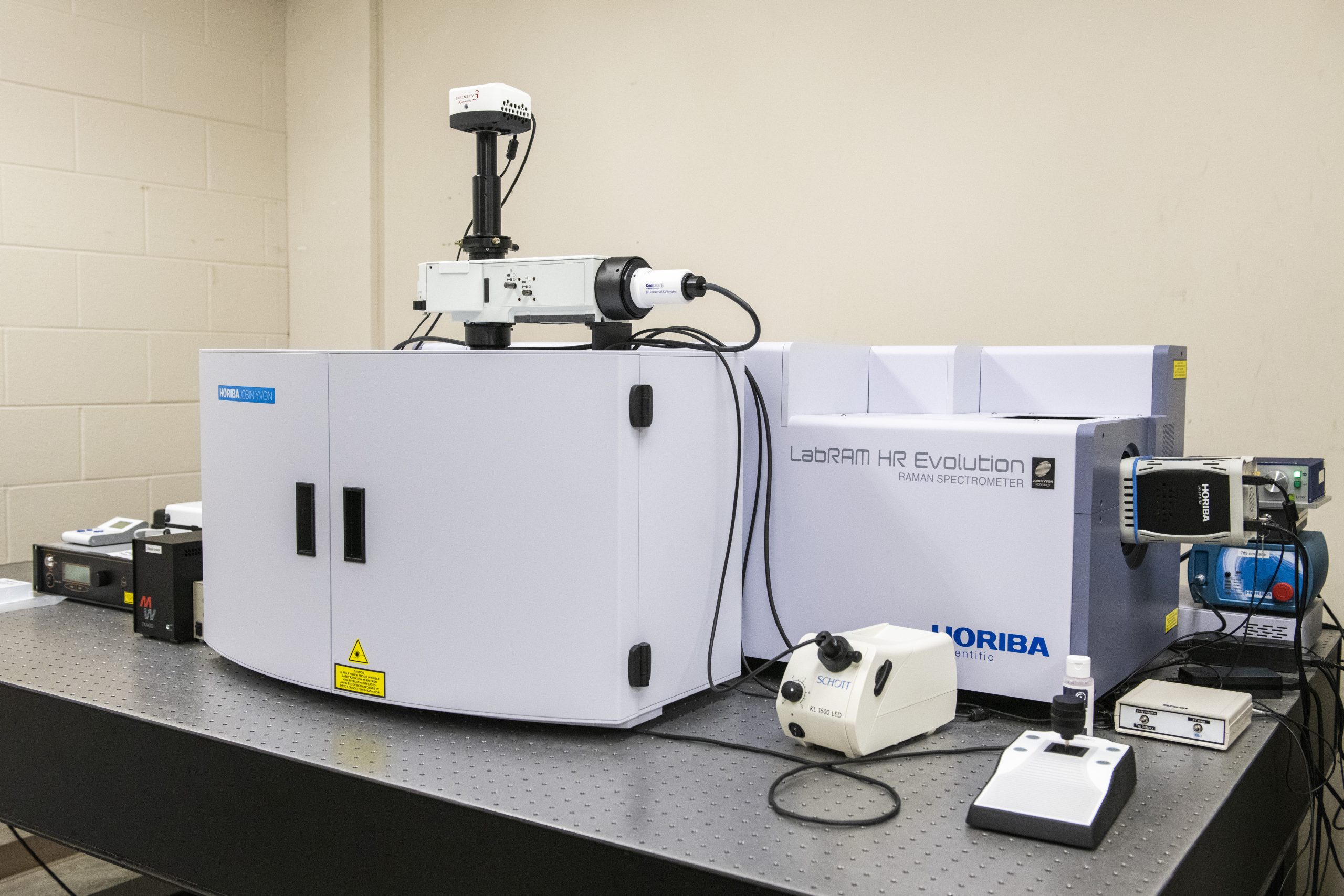
- White light laser
- FLIM and FRET capabilities
- Environmental chamber with heat and gas control
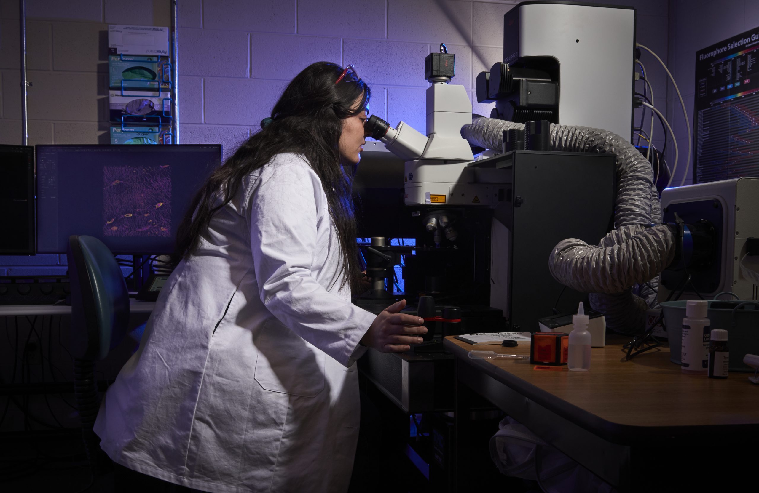
- Stimulated raman spectroscopy with CARS laser
- White light laser
- Environmental chamber with gas and temperature control
- Fully automated timelapse imaging
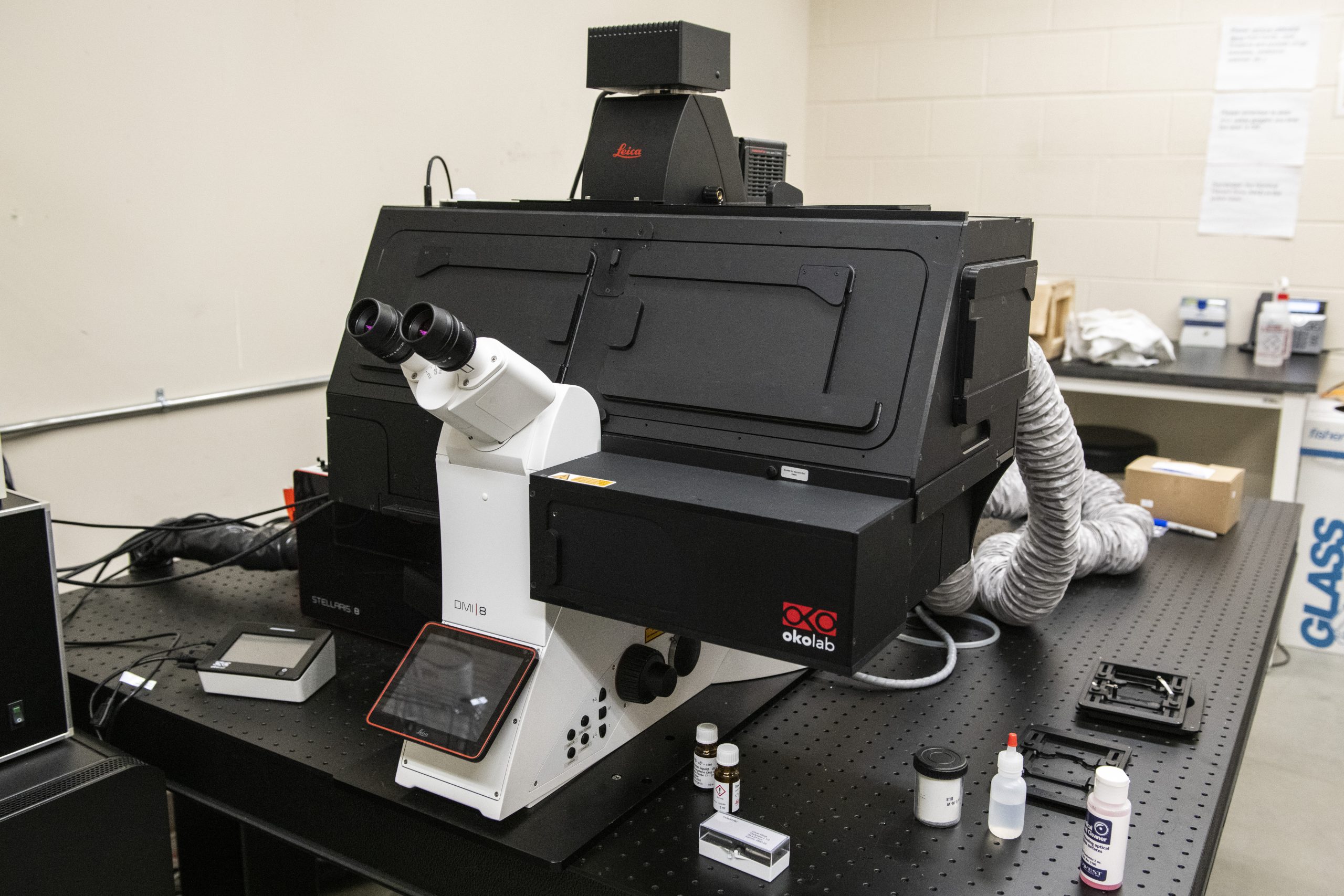
- Optical Tweezers
- Fast-mapping fluorescence capability with 450 mW 532 nm laser and a 100 mW 785 nm
laser
- Ultra-low frequency filters for Stokes and anti-Stokes measurements
- High-sensitivity, high-throughput imaging
- fully enclosed to maintain temperatures between ambient 5-50°C (± 0.1°C), humidity
(specifically designed to prevent condensation), and CO2/Air or hypoxic/hyperoxia conditions
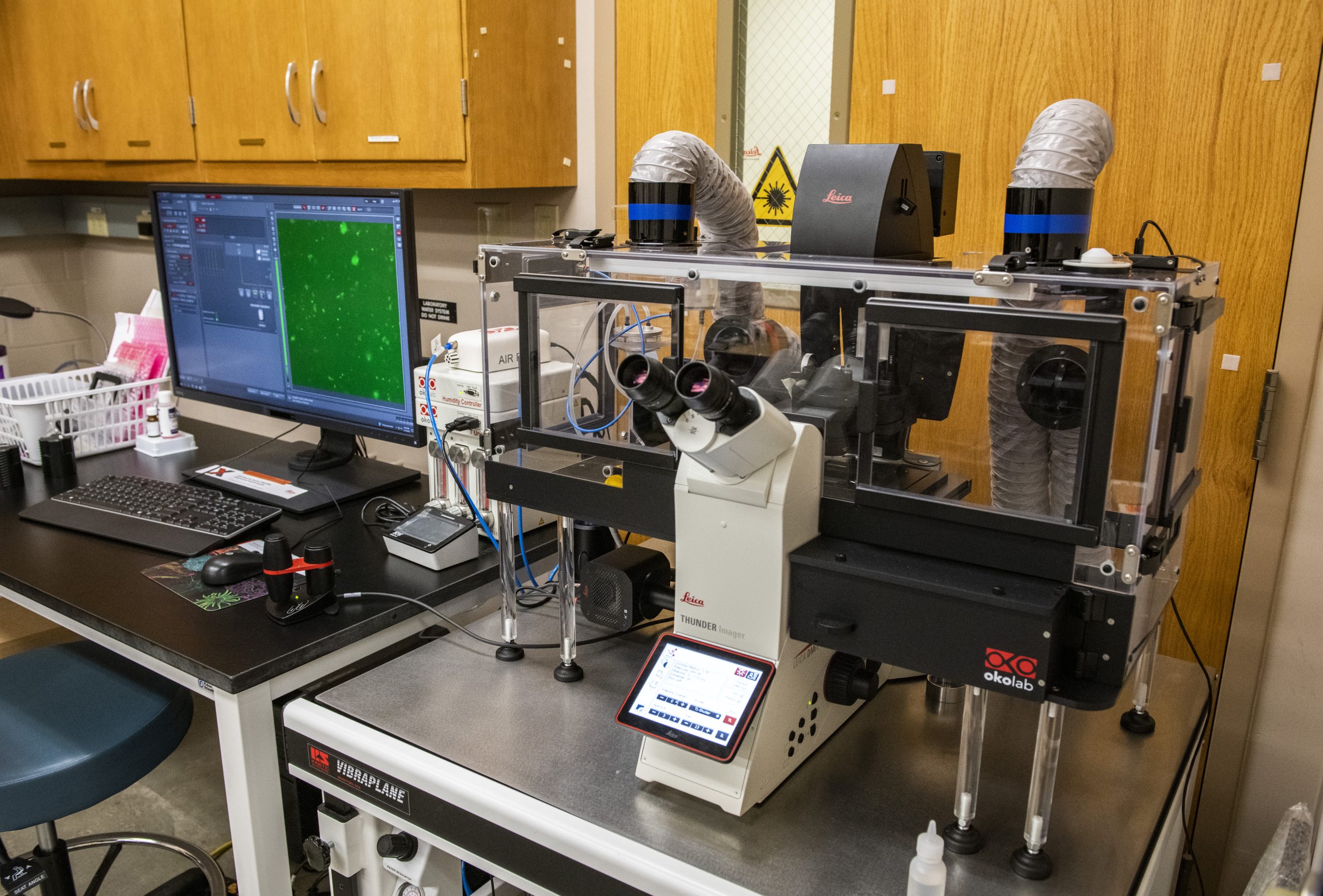
- Enables the precise isolation of cells or specific regions of interest
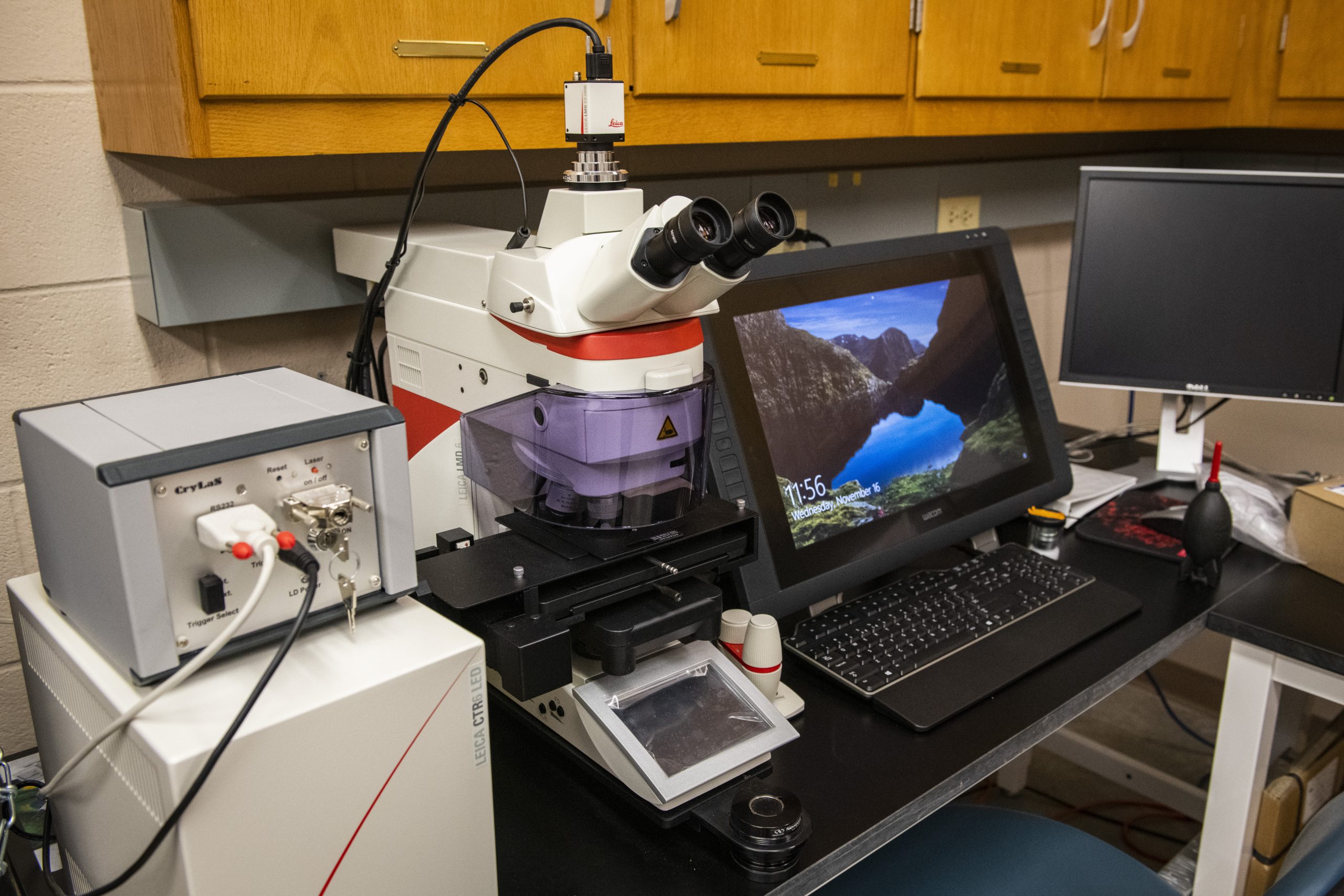
- UV laser for sample dissection
- Equipped with a color camera and fluorescence filter cubes (FITC, TRITC, DAPI)
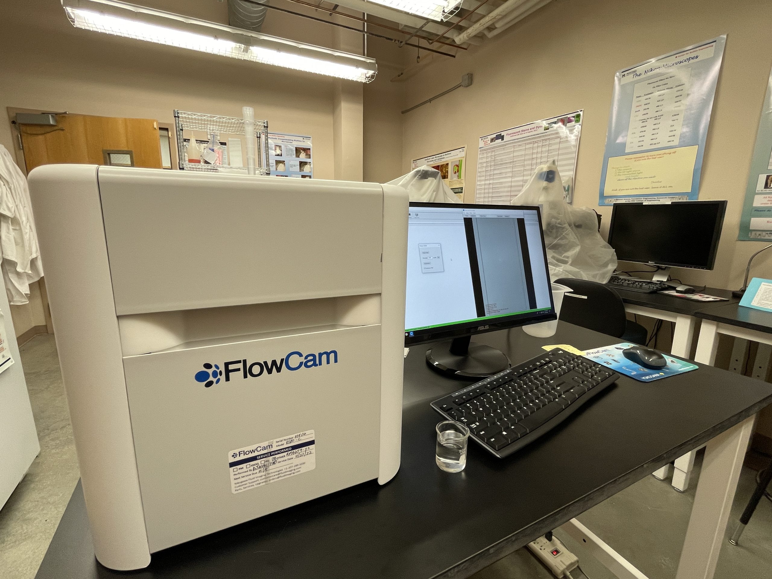
- Flow imaging microscopy system
- Combines the benefits of digital imaging flow cytometry, and microcopy into a single
platform
- Count passing particles in a liquid sample
- Characterize suspended particles in a size range of 2µm to 1mm
- Leica DFC3000G fluorescence camera
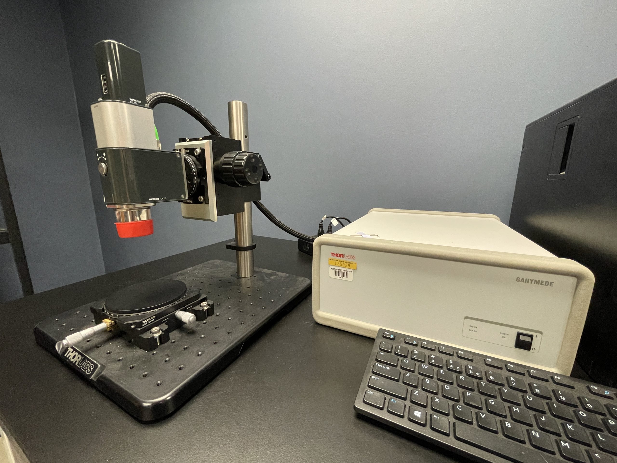
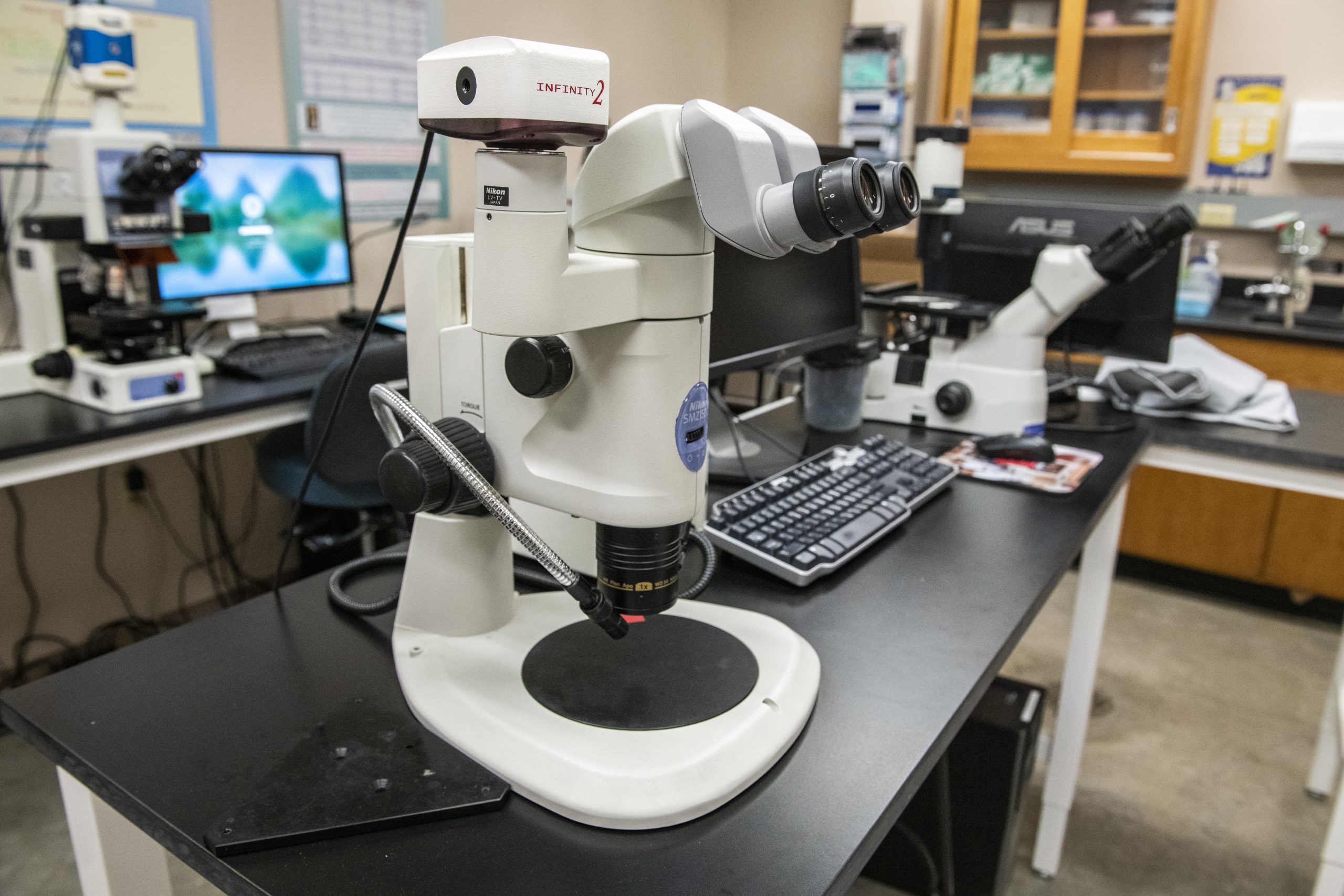
- Computer-controlled stereomicroscope
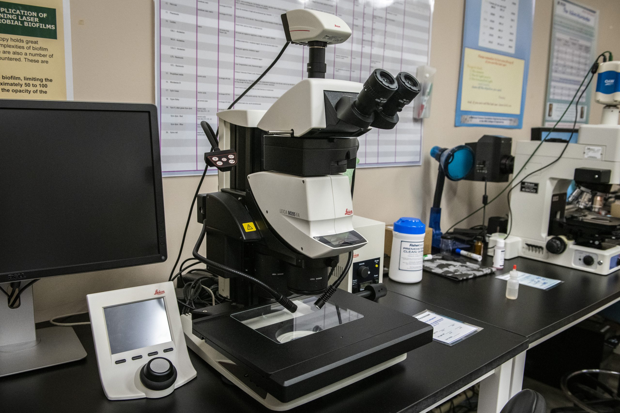
- Enables a stain-free, non-invasive, high resolution, real-time imaging of thick intact
samples
- Fully automated with constant color temperature
- Fully automated transmitted light- and fluorescence axis
- Motorized Z-focus
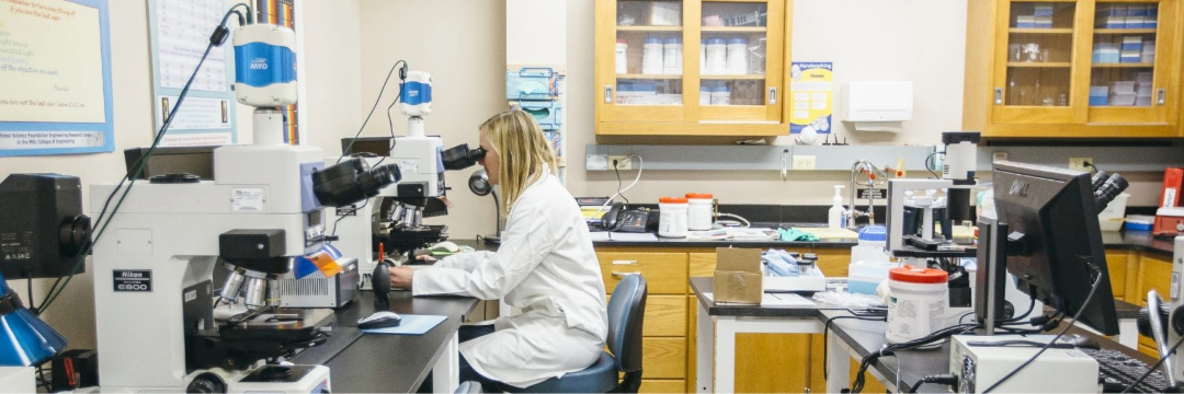
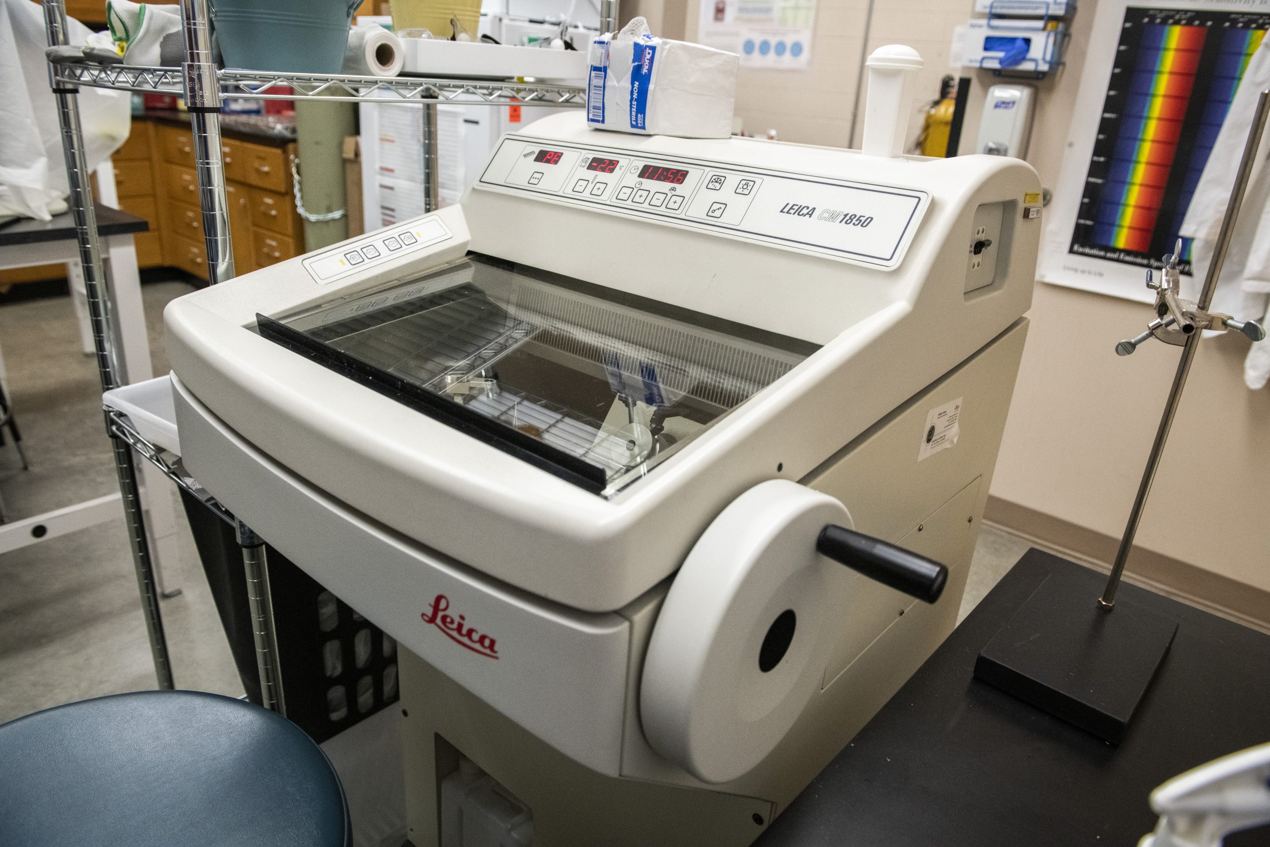
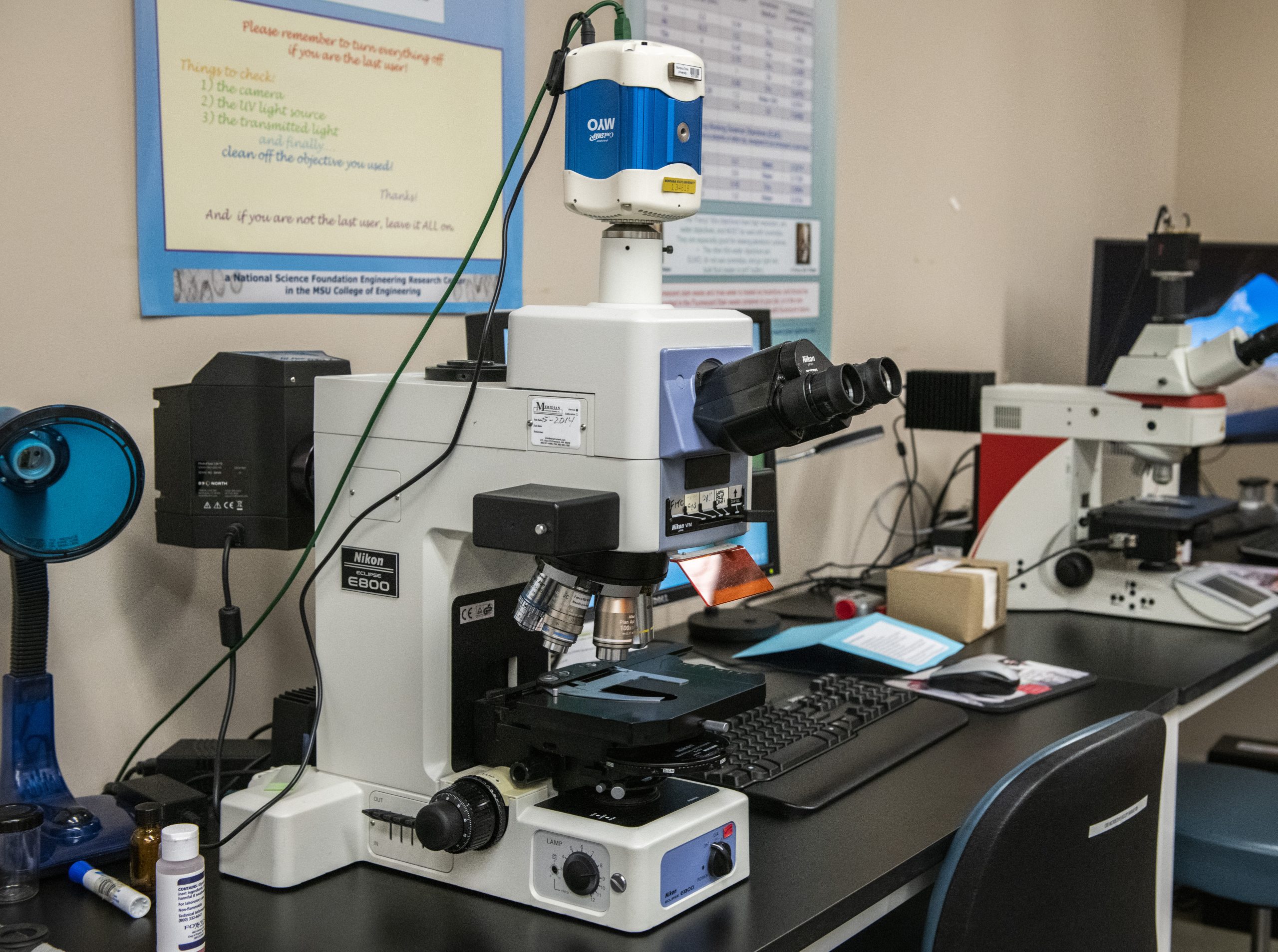
- Cryostat at -20C for thin-sectioning biofilms or tissues
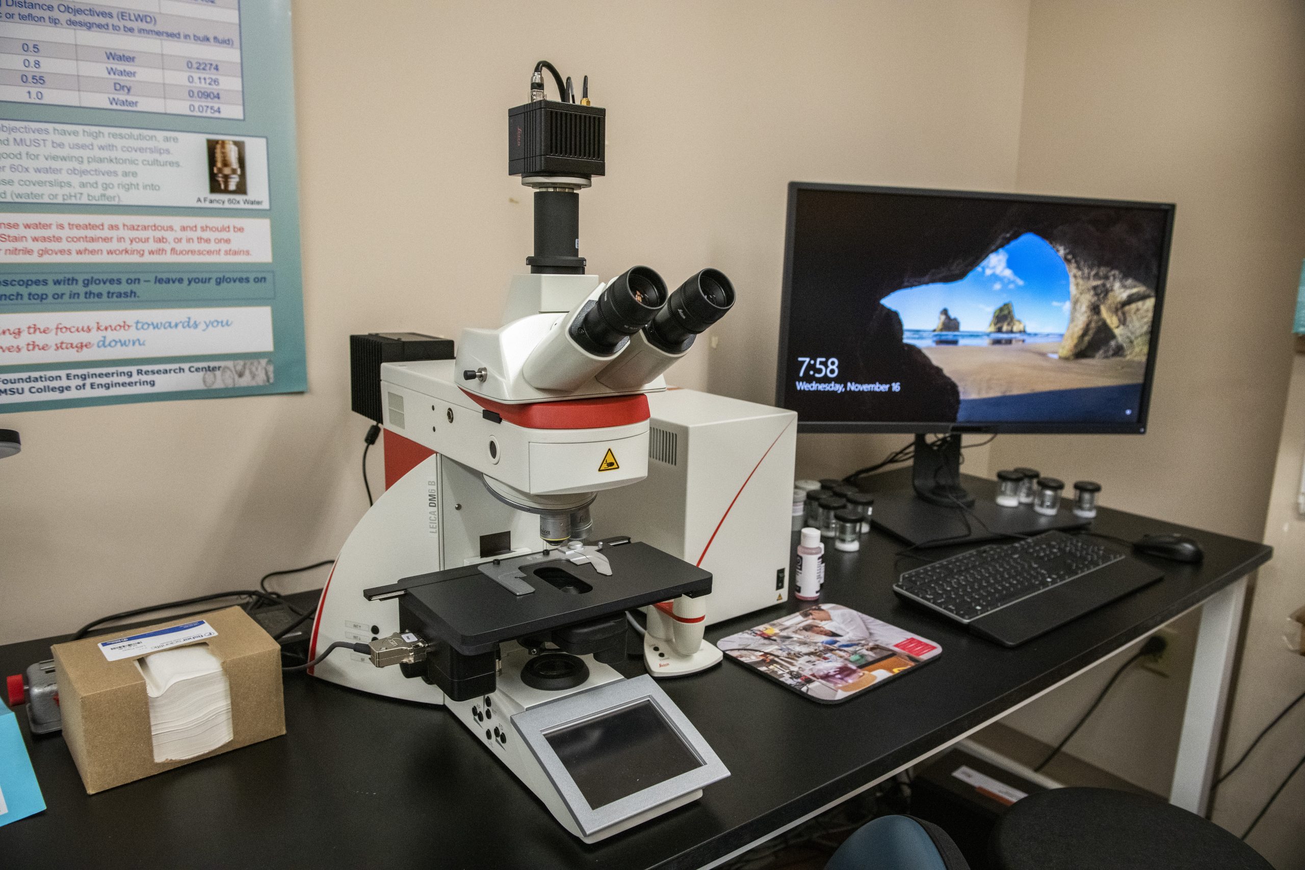
- Fluorescence filters
- Photometrics MYO cooled CCD camera
- Universal Imaging Corporation’s MetaVue software (v7.4.6)
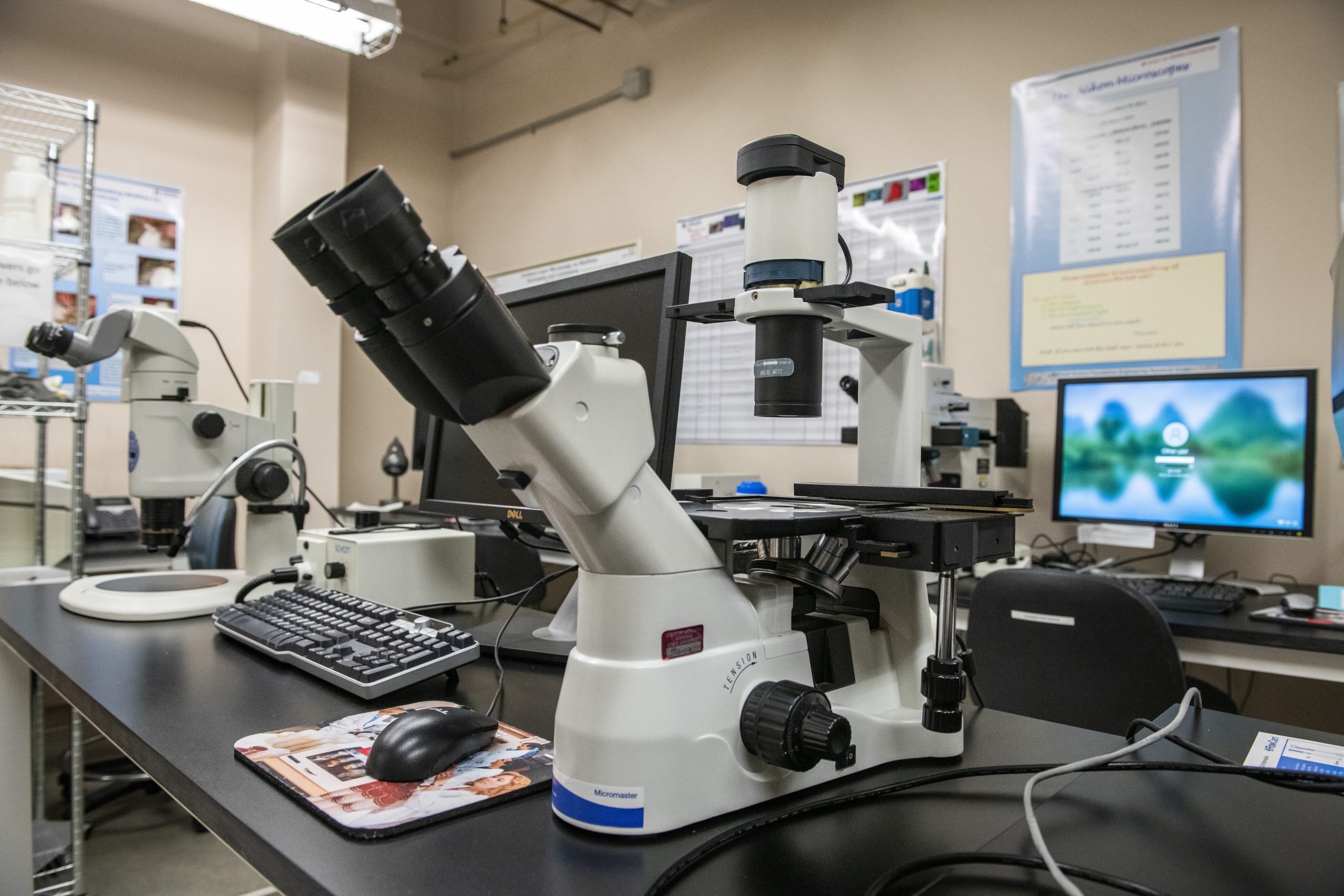
- Barrel zoom
- Equipped with a color camera
|